Proudly Part of Privia Health
Sports Medicine/Tendon/Bone-Joint Injury
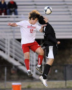
At Dominion Foot and Ankle Consultants we see many patients with sports-related and tendon injuries. Some of these injuries have acute onset such as tendon rupture or bone fracture, others are as a result of chronic overuse, meaning that they develop over weeks to months based on structural problems related to the foot or ankle, improper training technique, or simply overtraining without allowing adequate periods of recovery. Below are some examples of each.
Acute tendon injury:
Below is an example of acute Achilles tendon rupture, MR image and minimally-invasive repair through several small incisions. This creates less scarring and minimal disruption to blood flow to tendon, resulting in quicker healing and return to function than conventional, open approaches.
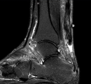
Achilles rupture on MRI
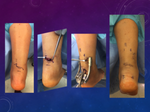
Minimally-invasive Achilles tendon repair
Chronic tendon injury:
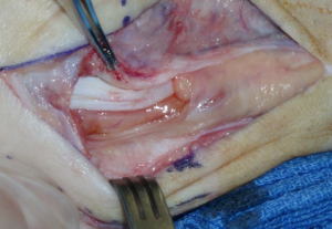
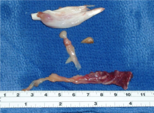
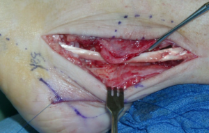
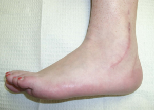
Below is an example of a peroneal tendon tear, patient had a history of ankle sprains but also a structural deformity of the heel which resulted in continual, low-grade injury of the peroneus brevis tendon (longitudinal tear). By the time they came to our care, a portion of the tendon was so damaged that portion along with peroneal tubercle had to be excised, and the remainder repaired, see images below:
|
Peroneus tendon tear |
Peroneus tendon tear resected |
Tendon repair/ tenodesis peroneus brevis to longus |
|
Healed tendon repair and calcaneal osteotomy (heel realignment) |
Improved heel alignment associated with original tendon tear |
Below is an example of a posterior ankle impingement syndrome (chronic pain in back of ankle) in a youth lacrosse player, an accessory (“extra”) bone is present and contributes to the problem, arthroscopic excision is accomplished to resolve issue.
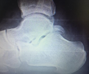
Large os trigonum
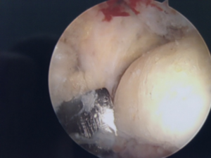
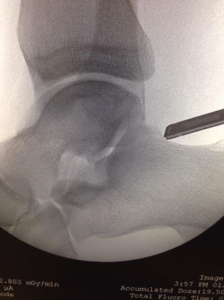
Arthroscopic removal of symptomatic os trigonum causing chronic pain
Below is an example of overuse injury in a 16-year old basketball player resulting in stress fracture of the navicular, plain x-rays were normal so a bone scan was obtained, see image with navicular “lighting up,” immobilization and non-weightbearing allowed complete healing and return to play.
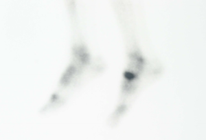
Navicular stress fracture on bone scan, x-rays were “normal”
